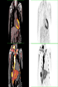
Renal Metastasis of Primary Lung Carcinoma is Associated with Death and Progression Predicted by F-18 FDG PET/CT
Renal Metastasis of Primary Lung Carcinoma is Associated with Death and Progression Predicted by F-18 FDG PET/CT
kidney metastasis, lung carcinoma, FDG, PET,
___
- 1. Longo, R., Jaud, C., Gehin, W., Hennequin, L., Bastien, C., Campitiello, M., Rozzi, A., & Plastino, F. (2020). Renal Metastases from a Nasal Cavity Mixed Squamous Cell and Adenoid Cystic Carcinoma: A Case Report. The American journal of case reports, 21, e919781. https://doi.org/10.12659/AJCR.919781
- 2. Hassoun, H., Alabed, Y. Z., Karls, S., Probst, S., & Laufer, J. (2016). 18F-FDG PET/CT Imaging of Bilateral Renal Metastasis of Breast Adenoid Cystic Carcinoma. Clinical nuclear medicine, 41(2), 148–149. https://doi.org/10.1097/RLU.0000000000001047
- 3. Basu, S., Ramani, S. K., & Lad, S. (2009). Unusual involvement of scalp and bilateral kidneys in an aggressive mediastinal diffuse large B cell lymphoma: documentation by FDG-PET imaging. Clinical nuclear medicine, 34(9), 638–641. https://doi.org/10.1097/RLU.0b013e3181b06abf
- 4. Bailey, J. E., Roubidoux, M. A., & Dunnick, N. R. (1998). Secondary renal neoplasms. Abdominal imaging, 23(3), 266–274. https://doi.org/10.1007/s002619900337
- 5. Ho, C. L., Chen, S., Ho, K. M., Chan, W. K., Leung, Y. L., Cheng, K. C., Wong, K. N., Cheung, M. K., & Wong, K. K. (2012). Dual-tracer PET/CT in renal angiomyolipoma and subtypes of renal cell carcinoma. Clinical nuclear medicine, 37(11), 1075–1082. https://doi.org/10.1097/RLU.0b013e318266cde2
- 6. Aras, M., Dede, F., Ones, T., Inanır, S., Erdil, T. Y., & Turoğlu, H. T. (2013). Is The Value of FDG PET/CT In Evaluating Renal Metastasis Underestimated? A Case Report And Review of The Literature. Molecular imaging and radionuclide therapy, 22(3), 109–112. https://doi.org/10.4274/Mirt.130
- 7. Zhao, Q., Dong, A., Yang, B., Wang, Y., & Zuo, C. (2017). FDG PET/CT in 2 Cases of Renal Metastasis From Esophageal Squamous Cell Carcinoma. Clinical nuclear medicine, 42(11), 896–898. https://doi.org/10.1097/RLU.0000000000001813
- 8. Bracken, R. B., Chica, G., Johnson, D. E., & Luna, M. (1979). Secondary renal neoplasms: an autopsy study. Southern medical journal, 72(7), 806–807. https://doi.org/10.1097/00007611-197907000-00013
- 9. Sánchez-Ortiz, R. F., Madsen, L. T., Bermejo, C. E., Wen, S., Shen, Y., Swanson, D. A., & Wood, C. G. (2004). A renal mass in the setting of a nonrenal malignancy: When is a renal tumor biopsy appropriate?. Cancer, 101(10), 2195–2201. https://doi.org/10.1002/cncr.20638
- 10. Kochhar, R., Brown, R. K., Wong, C. O., Dunnick, N. R., Frey, K. A., & Manoharan, P. (2010). Role of FDG PET/CT in imaging of renal lesions. Journal of medical imaging and radiation oncology, 54(4), 347–357. https://doi.org/10.1111/j.1754-9485.2010.02181.x
- 11. Nakagawa, T., Fujimura, T., Morikawa, T., Fukayama, M., Homma, Y., Ohtomo, K., & Momose, T. (2015). Preoperative evaluation of renal cell carcinoma by using 18F-FDG PET/CT. Clinical nuclear medicine, 40(12), 936–940. https://doi.org/10.1097/RLU.0000000000000875 12. Gündoğan, C., Çermik, T. F., Erkan, E., Yardimci, A. H., Behzatoğlu, K., Tatar, G., Okçu, O., & Toktaş, M. G. (2018). Role of contrast-enhanced 18F-FDG PET/CT imaging in the diagnosis and staging of renal tumors. Nuclear medicine communications, 39(12), 1174–1182. https://doi.org/10.1097/MNM.0000000000000915
- 13. Nakhoda, Z., Torigian, D. A., Saboury, B., Hofheinz, F., & Alavi, A. (2013). Assessment of the diagnostic performance of (18)F-FDG-PET/CT for detection and characterization of solid renal malignancies. Hellenic journal of nuclear medicine, 16(1), 19–24. https://doi.org/10.1967/s002449910067
- Başlangıç: 2021
- Yayıncı: Mersin Üniversitesi
F-18 FDG PET/CT in a Soft Tissue Tumor with Pericardial Metastasis
Gökçe YAVAN, Zehra Pınar KOÇ, Pelin Özcan KARA, Hamide SAYAR, Ayşe TÜRKMEN, Ahmet ÇELİK, Emel SEZER
Zehra Pınar KOÇ, Zeynep Selcan SAĞLAM, Pelin Özcan KARA, Gökçe YAVAN, Hasan Hüsnü YÜKSEK
PET-CT Imaging in Cancer of Unknown Primary in a Patient with Cranial Metastatic Mass
Pelin Özcan KARA, Zehra Pınar KOÇ, Adil GÜMÜŞ, Mahmut SÜLEYMANOĞLU
Zehra Pınar KOÇ, Pelin Özcan KARA, Vehbi ERÇOLAK, Yasemin YUYUCU KARABULUT
Pelin Özcan KARA, Zehra Pınar KOÇ, Mehmet ARSLAN, Mahmut SÜLEYMANOĞLU, Rabia KURUKAHVECİ
