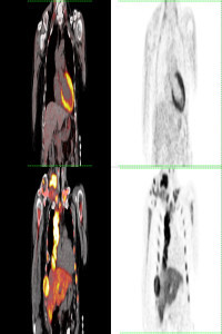
Bladder Crystal Formation shown as false positive cause in F-18 Fluorodeoxyglucose (FDG) Positron Emission Tomography/Computed Tomography (PET/CT)
Bladder Crystal Formation shown as false positive cause in F-18 Fluorodeoxyglucose (FDG) Positron Emission Tomography/Computed Tomography (PET/CT)
Crystal, False Positive FDG PET/CT,
___
- 1. Boudabbous S., Arditi D., Paulin E., Koessler T., Rougemont A.L., Montet X. Ossifying metaplasia of urothelial metastases: original case with review of the literature. BMC Med Imaging. 2015 ;15:30
- 2. Cormio L., Sanguedolce F., Massenio P., Di Fino G., Selvaggio O., Bufo P., et al. Osseous metaplasia within a urothelial bladder cancer nodal metastasis: a case report. Anal Quant Cytopathol Histpathol. 2014;36:117–9
- 3. Gladdy R.A., Qin L.X., Moraco N., Edgar M.A., Antonescu C.R., Alektiar K.M., et al. Do radiation-associated soft tissue sarcomas have the same prognosis as sporadic soft tissue sarcomas? J Clin Oncol. 2010;28:2064–9
- 4. Mantzarides M., Papathanassiou D., Bonardel G., et al. High-grade lymphoma of the bladder visualized on PET. Clin Nucl Med. 2005; 30:478-80
- 5. Treglia G., Bongiovanni M., Giovanella L. A rare case of small cell neuroendocrine carcinoma of the urinary bladder incidentally detected by F-18 FDG PET/CT. Endocrine. 2014; 45:156-157
- 6. Lin Y., Lu Y.Y., Wang H.Y., Tsai S.C., Lin W.Y. Accidental finding of bladder cancer in 99mTc methylene diphosphonate whole-body bone scan. Clin Nucl Med. 2013;38:643-5
- 7. Parekh A., Hagan I., Capaldi L., Lyburn I. Incidental Papillary Bladder Carcinoma on 18F-Fluoromethylcholine PET/CT Undertaken to Evaluate Prostate Malignancy. Clin Nucl Med. 2017 .;42:721-722
- 8. Osman M.M., Altinyay M.E., Abdelmalik A.G., Brickman T.M., Varvares M.A., Nguyen N.C. FDG PET/CT incidental diagnosis of a synchronous bladder cancer as a fourth malignancy in a patient with head and neck cancer. Clin Nucl Med. 2011;36:496-7
- 9. De Jong I.J., Pruim J., Elsinga P.H., et al. Visualisation of bladder cancer using (11)C-choline PET: first clinical experience. Eur J Nucl Med Mol Imaging. 2002;29:1283–1288
- 10. Tahara T., Ichiya Y., Kuwabara Y., et al. High [18F]-fluorodeoxyglucose uptake in abdominal abscesses: a PET study. J Comput Assist Tomogr. 1989;13: 829–831
- Başlangıç: 2021
- Yayıncı: Mersin Üniversitesi
Zehra Pınar KOÇ, Pınar Pelin ÖZCAN, Tahsin ÇOLAK, Gökçe YAVAN
Pınar Pelin ÖZCAN, Zehra Pınar KOÇ, Gökçe YAVAN, Adil GÜMÜŞ
Pınar Pelin ÖZCAN, Zehra Pınar KOÇ, Gökçe YAVAN, Vehbi ERÇOLAK
F-18 FDG PET/CT imaging in the Ameloblastoma Follow Up
Zehra Pınar KOÇ, Pınar Pelin ÖZCAN, Emel SEZER, Gökçe YAVAN
The Effect of Portable Gamma Camera in Radioguided Lesion Localization and The Patient Management
Zehra Pınar KOÇ, Pınar Pelin ÖZCAN, Yüksel BALCI, Mustafa BERKEŞOĞLU, Ferah TUNCEL DALOĞLU
Pınar Pelin ÖZCAN, Zehra Pınar KOÇ, Gökçe YAVAN, Mustafa Musa DİRLİK
