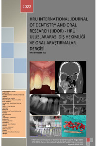
Examination of the Relationship of Dental İmplants in the Maxilla with the Nasopalatine Canal by Cone-Beam Computed Tomography
Examination of the Relationship of Dental İmplants in the Maxilla with the Nasopalatine Canal by Cone-Beam Computed Tomography
___
- 1. White SC, Pharoah Michael J. Oral Radiology: Principles Anf Interpretation: Elsevier; 2012.
- 2. Langland OE, Langlais RP. Early pioneers of oral and maxillofacial radiology. Oral Surg Oral Med Oral Pathol Oral Radiol Endod. 1995;80(5):496-511.
- 3. Fokas G, Vaughn VM, Scarfe WC, Bornstein MM. Accuracy of linear measurements on CBCT images related to presurgical implant treatment planning: a systematic review. Clin Oral Implants Res. 2018;29:393-415.
- 4. Özalp Ö, Tezerişener HA, Kocabalkan B, Büyükkaplan UŞ, Özarslan MM, Kaya GŞ, et al. Comparing the precision of panoramic radiography and conebeam computed tomography in avoiding anatomical structures critical to dental implant surgery: A retrospective study. Imaging Sci Dent. 2018;48(4):269-75.
- 5. Kaya Y, Sarikcioglu L. Sir Herbert Seddon (1903–1977) and his classification scheme for peripheral nerve injury.Childs Nerv Syst. 2015;31(2):177-80.
- 6. Pamukçu U, İspir NG, Alkurt MT, Altunkaynak B, Peker İ. The Retrospective Assessment of Complications in Dental Implants Via ConeBeam Computed Tomography. Selcuk Dental Journal. 2021;8(2):367-71.
- 7. Gaêta-Araujo H, Oliveira-Santos N, Mancini AXM, Oliveira ML, OliveiraSantos C. Retrospective assessment of dental implant-related perforations of relevant anatomical structures and inadequate spacing between implants/teeth using cone-beam computed tomography.Clin Oral Investig 2020;24(9):3281-8.
- 8. Liaw K, Delfini RH, Abrahams JJ. Dental Implant Complications. Semin Ultrasound CT MR. 2015; 36: 427-33.
- 9. Genç T. Radiological Evaluation of Anatomical Structures and Variations in Maxilla and Mandible Before Dental Implant Treatment. 2014.
- 10. Hakbilen S, Mağat G. Nazopalatin kanal ve klinik önemi: Derleme. Selcuk Dental Journal. 2019;6(1):91-7.
- 11. Alkanderi A, Al Sakka Y, Koticha T, Li J, Masood F, Suárez‐López del Amo F. Incidence of nasopalatine canal perforation in relation to virtual implant placement: A cone beam computed tomography study. Clin Implant Dent Relat Res. 2020;22(1):77-83.
- 12. Cavallaro J, Tsuji S, Chiu T-S, Greenstein G. Management of the Nasopalatine Canal and Foramen Associated With Dental Implant Therapy. Compend Contin Educ Dent (Jamesburg, NJ: 1995). 2016;38(6):367-72
- Başlangıç: 2021
- Yayıncı: Harran Üniversitesi
Muhammed DEMİR, Fatih TULUMBACI
Mehmet Emin DOĞAN, Eda Didem YALÇIN
Endokron Restorasyonlar ve Endokron Restorasyonlarda Kullanilan Materyaller
Esengül SEZGİN, Yakup KANTACI, Burcu ÜSTÜN
Evaluation of Delayed Splint Removal and Root Resorption due to Pandemic: Case Report
Mesut KAYGUSUZ, Mehmet Sinan DOĞAN, Ayesha TAHA
Association of consanguineous marriages with severe early childhood caries: a pilot study
Ahmet ARAS, Mehmet Sinan DOĞAN, Şemsettin YILDIZ, Muhammed DEMİR
An In Vitro Microleakage Evaluation of a New Cold Flowable Gutta-Percha Based Sealer
Oculo-Dento-Digital Dysplasia (Oddd), Report of a Case
Pamela ARMİ, Roberta D’AVENİA, Cristina VİLLANACCİ, Francisco Cammarata SCALİSİ, Michele CALLEA
Çocuklarda Endodontik Enfeksiyonlara Bağlı Antibiyotik Kullanımı
Gizem KARAGÖZ DOĞAN, İsmet Rezani TOPTANCI
