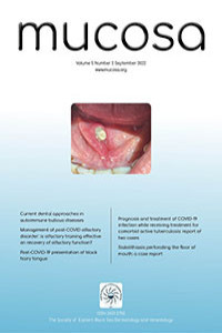
Ağız tabanını perfore eden tükürük bezi taşı: Olgu sunumu
Sialolithiasis, tükürük bezinin en sık görülen hastalığıdır. En sık submandibular bezde bulunur, ancak daha az sıklıkla parotis ve sublingual bezlerde ortaya çıkar. Submandibular bez parotis bezine göre sialolitiazise daha yatkındır. Çünkü Wharton kanalı daha geniş ve daha uzundur, milohyoid kasın arka sınırında yerçekimine karşı açılanma yapar ve submandibular bez sekresyonu daha alkali, müsinöz, kalsiyum ve fosfattan daha zengin ve daha yavaş akış hızına sahiptir. Bu olgu sunumunda ağız tabanı mukozası bir sialolit tarafından perfore edilmiş bir hasta sunuldu. Sialolitler nadiren görülmekle birlikte tekrarlayan ciddi enfeksiyonlara ve hastalarda ağrıya neden olarak yaşam kalitesini olumsuz etkiler. Bu nedenle klinisyen ağız tabanında yabancı cisim hissi veya çene altında yemekle ilişkili şişlik olması durumunda submandibular tükürük bezi taşını aklına getirmelidir.
Anahtar Kelimeler:
submandibular bez, sialolitler, sialolithiasis
Sialolithiasis perforating the floor of mouth: a case report
Sialolithiasis is the most common disease of the salivary gland. It is most commonly found in the submandibular gland, but less frequently in the parotid and sublingual glands. The submandibular gland is more prone to sialolithiasis than the parotid gland, because Wharton’s canal is wider and longer, it angulates against gravity at the posterior border of the mylohyoid muscle and submandibular gland secretion is more alkaline, mucinous, richer in calcium and phosphate and slower flow rate. In this case report, a patient whose floor of the mouth mucosa was perforated by a sialolith was presented. Although sialoliths are infrequently seen, they cause severe recurrent infections and pain in patients and adversely affect the quality of life. Therefore, the clinician should consider submandibular sialolithiasis in case of foreign body sensation in the floor of the mouth or swelling under the chin associated with a meal.
Keywords:
sialolithiasis, submandibular gland, sialolithotomys,
___
- Teymoortash A, Buck P, Jepsen H, Werner JA. Sialolith crystals localized intraglandularly and in the Wharton’s duct of the human submandibular gland: an X-ray diffraction analysis. Arch Oral Biol 2003;48:233-6.
- Lustmann J, Regev E, Melamed Y. Sialolithiasis A survey on 245 patients and a review of the literature. Int J Oral Max Surg 1990;19:135-8.
- Oteri G, Procopio RM, Cicciu M. Giant salivary gland calculi (GSGC): report of two cases. Open Dent J 2011;5:90-5.
- Lim EH, Nadarajah S, Mohamad I. Giant submandibular calculus eroding oral cavity mucosa. Oman Medical J 2017;32:432-5.
- Erdogan O, Ozcan C, Ismi O, Gur H, Vayisoglu Y, Gorur K. Effectiveness of sialendoscopy on the symptoms of chronic obstructive sialadenitis and patient satisfaction degree. J Stomatology Oral Maxillofac Surg 2022;123:314-9.
- Parkar MI, Vora MM, Bhanushali DH. A large sialolith perforating the Wharton’s duct: review of literature and a case report. J Maxillofac Oral Surg 2012;11:477-82.
- Arslan S, Vuralkan E, Cobanoglu B, Arslan A, Ural. A. Giant sialolith of submandibular gland: report of a case. J Surg Case Rep 2015;4:1-3.
- Dam VSKE, Umbaik NAM, Lazim NM. Self-Extrusion of unusual size submandibular sialolith: a case report. Gazi Medical J 2022;33:69-72.
- ISSN: 2651-2750
- Yayın Aralığı: Yılda 3 Sayı
- Başlangıç: 2018
- Yayıncı: Doğu Karadeniz Deri ve Zührevi Hastalıklar Derneği
