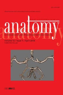
Occipital emissary foramina in human skulls: review of literature and proposal of a classification scheme of the occipital venous anastomoses in the posterior cranial fossa
___
- Federative Committee of Anatomical Terminology (FCAT). Terminologia anatomica: International anatomical terminology. Stuttgart: Georg Thieme Verlag; 1998. p. 1-292.
- Clemente CD (ed). Anatomy of the human body. 30th ed. Philadelphia: Lea and Febiger; 1985. p. 1-1676.
- Williams PL, Bannister LH, Berry MM, Collins P, Dyson M, Dussek JE, Ferguson MWJ (eds). Gray’s anatomy. 38th ed. Edinburgh: Churchill Livingstone; 1995. p. 1-2092.
- Standring S (ed) Gray’s anatomy: The anatomical basis of clinical practice. 41st ed. London: Elsevier; 2016. p. 1-1562.
- Boyd GI. The emissary foramina of the cranium in man and the anthropoids. J Anat 1930;65:108-21.
- Kadanoff D, Mutafov S. The human skull in a medico-anthropological aspect: form, dimensions and variability. Sofia: Prof. Marin Drinov Academic Publishing House; 1984. p. 121.
- Sharma PK, Malhotra VK, Tewari SP. Emissary occipital foramen. Anat Anz 1986;162:297-8.
- Premsagar IC, Lakhtakia PK, Bisaria KK. Occipital emissary foramen in Indian skulls. J Anat 1990;173:187-8.
- Gözil R, Kadioglu D, Calgüner E. Occipital emissary foramen in skulls from Central Anatolia. Acta Anat (Basel)1995;153:325-6.
- Hossain SMA, Rahman L, Karim M. Occipital emissary foramen in Bangladeshi skulls. Pakistan Journal of Medical Sciences 2001;17:156-8.
- Louis RG Jr, Loukas M, Wartmann CT, Tubbs RS, Apaydin N, Gupta AA, Spentzouris G, Ysique JR. Clinical anatomy of the mastoid and occipital emissary veins in a large series. Surg Radiol Anat 2009;31:139-44.
- Murlimanju BV, Prabhu LV, Pai MM, Jaffar M, Saralaya VV, Tonse M, Prameela MD. Occipital emissary foramina in human skulls: an anatomical investigation with reference to surgical anatomy of emissary veins. Turk Neurosurg 2011;21:36-8.
- Singhal S, Ravindarath R. Occipital emissary foramina in South Indian modern human skulls. International Scholarly Research Notices (ISRN) Anatomy 2013:1-4.
- Cakmak PG, Ufuk F, Yagci AB, Sagtas E, Arslan M. Emissary veins prevalence and evaluation of the relationship between dural venous sinus anatomic variations with posterior fossa emissary veins: MR study. Radiol Med 2019;124:620-7.
- Hedjoudje A, Piveteau A, Gonzalez-Campo C, Moghekar A, Gailloud P, San Millán D. The Occipital emissary vein: A possible marker for pseudotumor cerebri. Am J Neuroradiol 2019;40:973-8.
- Mohsenipour I, Goldhahn W-E, Fischer J, Platzer W, Pomaroli A. Approaches in neurosurgery: Central and peripheral nervous system. Stuttgart: Georg Thieme Verlag; 1994. p. 107-25.
- San Millán Ruíz D, Gailloud P, Rüfenacht DA, Delavelle J, Henry F, Fasel JH. The craniocervical venous system in relation to cerebral venous drainage. Am J Neuroradiol 2002;23:1500-8.
- Okudera T, Huang YP, Ohta T, Yokota A, Nakamura Y, Maehara F, Utsunomiya H, Uemura K, Fukasawa H. Development of posterior fossa dural sinuses, emissary veins, and jugular bulb: morphological and radiologic study. Am J Neuroradiol 1994;15:1871-83.
- ISSN: 1307-8798
- Yayın Aralığı: 3
- Başlangıç: 2007
- Yayıncı: Deomed Publishing
Bilateral atresia of the external acoustic meatus: a case report
Mehmet ÖZTÜRK, Nadire ÜNVER DOĞAN, Zeliha FAZLIOĞULLARI, Ekrem SOLMAZ, Betül SEVİNDİK
Öznur ÖZALP, Hande SALIM, Busehan BİLGİN, Serra ÖZTÜRK, Merve SARIKAYA DOĞAN, Mehmet Berke GÖZTEPE, Engin ÇALGÜNER, Muzaffer SİNDEL, ALPER SİNDEL
Ivan Vasilyevich GAVORONSKİY, Ivan Antonovich LABETOV, Gleb Valerevich KOVALEV, Gennadii Ivanovich NİCİPORUK, Nikita Dmitrievich KUBİN, Dmitry Dmitrievich SHKARUPA
Evaluation of sternal morphology according to age and sex with multidetector computerized tomography
Güneş BOLATLI, Nadire ÜNVER DOĞAN, Mustafa KOPLAY, Zeliha FAZLIOGULLARI, Ahmet Kagan KARABULUT
Morphometry of the internal capsule on MR images in adult healthy individuals
Çiğdem ÖZER GÖKASLAN, Tolga ERTEKİN, Abdülkadir BİLİR, Ozan TURAMANLAR, Erdal HORATA, Hazal EMEKSİZ
Didem DÖNMEZ AYDIN, Enis ULUÇAM, Selman ÇIKMAZ, Menekşe KARAHAN, Ali YILMAZ, Muhammed PARLAK, Ayşe Zeynep YILMAZER KAYATEKİN
Ekrem SOLMAZ, MEHMET ÖZTÜRK, Zeliha FAZLIOGULLARI, Betül SEVİNDİK, Nadire DOĞAN ÜNVER
